Steps required to create a Virtual Reality experience through the human body.
by Suzanne Martin
Why is it used? VR offers invaluable educational advantages. It is ideal for showing clients products that are otherwise too expensive to physically demonstrate. The 3-D sensory impact is unmistakable
Steps required to create a Virtual Reality experience through the human body.
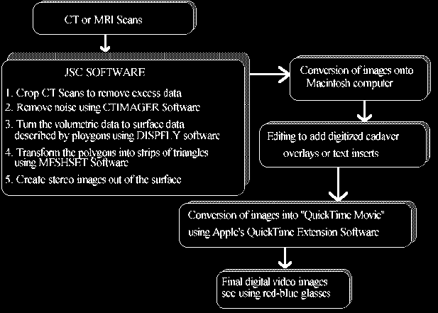
Viewing the final 3-D images was done on a Macintosh IIcx computer with 16M ram. A Mac was chosen because it is relatively affordable, it has widespread use in school systems, it is the leading engine of desktop multi-media, and there is a wide variety of software and hardware available for this task.
The VR movie can be stored either on a hard drive or transferred onto video tape and viewed through red-blue glasses. It can also be viewed using a VR Head-Mounted Display (HMD) or Binocular Omni Orientational Monitors (BOOM system). The final images can be stored on CD-ROM or laser disc.
A limitation due to the high resolution of the body images prohibits the observer from "flying" through the human body at will. Only the technology to create prescribed "fly throughs" is currently available. The technology is available for full interactive virtual reality experience with less data-intense applications, i.e. with applications requiring less resolution.
The data files created at the Medical Branch were then transferred to JSC IGOAL (Integrated Graphics, Operations, and Analysis Laboratory). There the scans were cropped to eliminate as much extraneous data as possible without losing any critical information. IGOAL developed a tool called "ctimager" which used thresholding to remove unwanted noise and extraneous data from each slice.
The filtering process typically consisted of thresholding the data to eliminate most of the noise. A low pass filter was used to minimize the high-frequency noise that would produce an irregular bumpy surface when input to the algorithm. This process produced a relatively smooth surface that approximated the scanned specimen and reduced the number of noise generated polygons. A unique filter was created for the heart data which only smoothed the data between scans, no other filtering was needed.
Due to the large number of slices in both the heart and skull data sets, several models were made, each of which represented a small number of slices. A meshing algorithm, "meshit", was developed to improve the display performance. This algorithm converted the raw collection of triangles into efficient strips. An average of over 100 triangles composed each triangle strip.
The Dartmouth Medical School in Hanover, N.H. has created computational models of the human face and lower extremities to examine the effects and outcomes of surgical procedures.
Greenleaf Medical Systems in Palo Alto, CA has created 'EVAL' and 'Glove Talker'. 'Eval' uses sensor-lined data gloves and data suits to obtain range-of-motion and strength measurements of injured and disabled patients. The 'Glove Talker' is a data glove sign-language device used for rehabilitation which allows someone without voice (stroke or cerebral palsy patient) to make gestures the computer understands. Using HMD, the patient can relearn how to open a door, walk, point or turn around in space.
Preliminary results have shown that a high resolution model can be developed using this type of imaging data. In order to maintain the goal of high quality VR imaging, some problems which were caused by the amount of data needed to deal with frame-by-frame sequencing of the prescribed fly-throughs had to be overcom. Alternative hardware and software solutions are being explored to alleviate this problem.
Another problem has been the technology for the HMD display systems. The LCD displays do not have the resolution needed to maintain a high quality VR experience. The CRT displays are reaching the resolution needed however the cost is prohibitive for multiple education platforms.
Surgical simulations may become routine especially to rehearse strategies for intricate and rare operations.
Porter, Stephen, "Virtual Reality", Computer Graphics World, (March, 1992), 42-54.
Sprague, Laurie A., Bell, Brad, Sullivan, Tim, and Voss, Mark, "Virtural Reality In Medical Education and Assessment", Technology 2003, December 1993.
![[Return to CS563 '95 talks list]](/images/back.gif)